What Is Medical Image Analysis?
Medical image analysis is the process of extracting meaningful information from medical images, often using computational methods. Some of the tasks for medical image analysis are visualization and exploration of 2D images and 3D volumes, segmentation, classification, registration, and 3D reconstruction of image data. The images for this analysis can be obtained from medical imaging modalities such as x-ray (2D and 3D), ultrasound, computed tomography (CT), magnetic resonance imaging (MRI), nuclear imaging (PET and SPECT), and microscopy.

You can load and display multimodal medical images from PET and CT using Medical Imaging Toolbox. (See MATLAB documentation.)
Medical image analysis can involve tasks such as counting and identifying cells in a microscopy image. For example, you can analyze and detect cancerous anomalies in the cells. For repetitive or subjective tasks, computational approaches can be used to automate or streamline medical image analysis and remove inconsistencies due to human error. With computational analysis, you can segment tumor tissues from necrosis or measure oxygen saturation in blood vessels.

Ten high-resolution blocks generated from the original multiresolution image of tumor tissue. MATLAB enables you to do block processing on very large images too big for memory and train deep learning networks on this data. (See code.)
By applying medical image analysis techniques, you can reconstruct a 3D representation from MRI images for calculating organ functions and other diagnostic measures.

You can segment and analyze brain MRI scans using the Medical Image Labeler app and MONAI in Medical Imaging Toolbox. (See code.)
Medical image analysis algorithms can be applied to large amounts of data, such as digital health data collected from wearable devices. You can use the algorithms to manage illnesses and health risks as well as promote health and well-being.
Medical Image Analysis with MATLAB
The MATLAB® development environment has built-in analysis and data access functionality for building algorithms for medical image analysis. With MATLAB, you can:
- Parse, load, visualize, and process medical file formats such as DICOM, NIfTI, and NRRD
- Visualize and explore 2D images and 3D volumes
- Use preprocessing techniques such as denoising, filtering, and augmentation
- Use deformable and nondeformable registration functions and apps to align multiple images and volumes
- Process very large multiresolution and high-resolution images
- Simplify medical image analysis tasks with built-in image labeling and segmentation algorithms and apps
- Create, modify, or transfer deep learning networks
In MATLAB, you can explore 2D images and 3D volumes using the Medical Image Labeler app. For example, you can load an MRI study of the human brain into the Medical Image Labeler, visualize the brain, label the individual areas, segment them, and analyze them for abnormalities.

The 3D volume data of a brain segmented using MONAI in the Medical Image Labeler app in Medical Imaging Toolbox. (See code.)
In digital pathology, whole tissue slides are imaged and digitized. The resulting whole slide images (WSIs) have extremely high resolution. Reading WSIs is a challenge because the images cannot be loaded into memory and therefore require out-of-core image processing techniques. MATLAB bigimage objects can store and process this type of large multiresolution image. Once the image is loaded, you can segment the cells using tools such as Cellpose.

With Medical Imaging Toolbox, you can perform block processing and use Cellpose to segment images, such as this image of a lymph node containing tumor tissue. (See code.)
MATLAB also includes apps for registration. For example, you can use the interactive Medical Registration Estimator app and Registration Estimator app to align 3D and 2D data, respectively.

Aligning multiple volumes using the Medical Registration Estimator app in Medical Imaging Toolbox. (See code.)
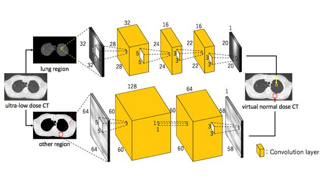
With MATLAB, you can also use deep learning methods to denoise, enhance, and segment 2D images and 3D volumes. You can design and train neural networks or use pretrained networks.

Segmented tumor in brain tissue using MATLAB with labeled ground truth (left) and network prediction (right). (See code.)
Examples and How To
Videos and Articles
Examples
Software Reference
See also: biological sciences, biotech and pharmaceutical, medical devices, image processing and computer vision

