Segmentation of WBC in microscopy images
版本 1.0.0 (1.8 MB) 作者:
Abhinav Gadge
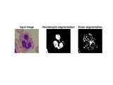
Stain deconvolution followed by threshold based segmentation is used.
Shape and size detection for WBC in blood is necessary for ensuring the ability to eliminate or control the infections or any inflammatory problems in the body.
WBC are of five types, subdivided into two parts,
1)Granulocytes: neutrophils , basophils, eosinophils
2)Agranulocytes: lymphocytes, monocyts.
The nucleus of WBC contains DNA and RNA that are stained by hematoxyline in Purple/blue color and the granules in cytoplasm are stained by eosine in pink color. As agranulocytes does not have granules in cytoplasm we won’t see eosine stained image for them. The function deconv is separating out the hematoxyline stained region and eosin stained region in different output images. The function segmentation can be used on this images to segment out nucleus and cytoplasmics granules.
Approach:
- Input RGB image is first deconvoluted to H and E stained Image.
- Find threshold for each output deconvolved image.
- Plot histogram representing two regions after segmentation.
- Apply segmentation based on threshold values.
- Output image is binary.
To see the results download this file, some output images are provided.
The segmentation can be done as follows.
InputImg = imread('ALL_IDB1\ALL_IDB1\im\Im061_1.jpg');
deconimg = deconv(InputImg);
H=deconimg(:,:,1);
E=deconimg(:,:,2);
[Sh,Hh,Th,Lh]=segmentation(H);
[Se,He,Te,Le]=segmentation(E);
引用格式
Abhinav Gadge (2024). Segmentation of WBC in microscopy images (https://www.mathworks.com/matlabcentral/fileexchange/102329-segmentation-of-wbc-in-microscopy-images), MATLAB Central File Exchange. 检索时间: .
MATLAB 版本兼容性
创建方式
R2021b
兼容任何版本
平台兼容性
Windows macOS Linux标签
Community Treasure Hunt
Find the treasures in MATLAB Central and discover how the community can help you!
Start Hunting!functions_output/function
functions_output/function
| 版本 | 已发布 | 发行说明 | |
|---|---|---|---|
| 1.0.0 |

